Oxford scientists watch a virus shell build itself, molecule by molecule
The team followed the assembly of a virus-like particle one molecule at a time. The study, published in Nature on 16 September 2026, is basic research: there is no drug yet, but it sheds light on a process future vaccines and antivirals could exploit.
Five protein pieces close into a pentagon-shaped ring, and from then on everything speeds up. It is one of the moments a team at the University of Oxford was able to see by following, molecule by molecule, how the shell of a virus-like particle puts itself together. The study was published in Nature on 16 September 2026. It is basic research: there is no drug yet, but it helps explain a process that future antivirals could try to disrupt.
Weighing a particle as it grows
Viruses keep their genetic material inside a protein shell, the capsid, which assembles itself from many pieces. Seeing the steps in between has been very hard, because those intermediate structures are scarce and short-lived.
Philipp Kukura’s group used a lab-engineered particle made of 60 protein units. They combined mass photometry, a technique developed at Oxford that measures the mass of single molecules from the light they scatter, with a new way of trapping each particle. That let them “weigh” it again and again as new pieces joined. “This method lets us study the assembly process directly, we just take a video!” says PhD student Dan Loewenthal, co-first author.
A maze where only the right turns stick
The team compares assembly to finding a way through a maze in which only the productive turns lock into place. At first the pieces bind weakly and reversibly: if an arrangement doesn’t work, they come apart and try again. When they form certain closed structures, such as that first pentagon, their multiple connections make them stable. Those few waypoints steer the process towards the finished particle, even though there are thousands of possible routes.
“The weak interactions give the system room to make mistakes,” says co-lead author Roi Asor. “That combination of trial and error followed by locking in successful structures is what makes the process so reliable.”
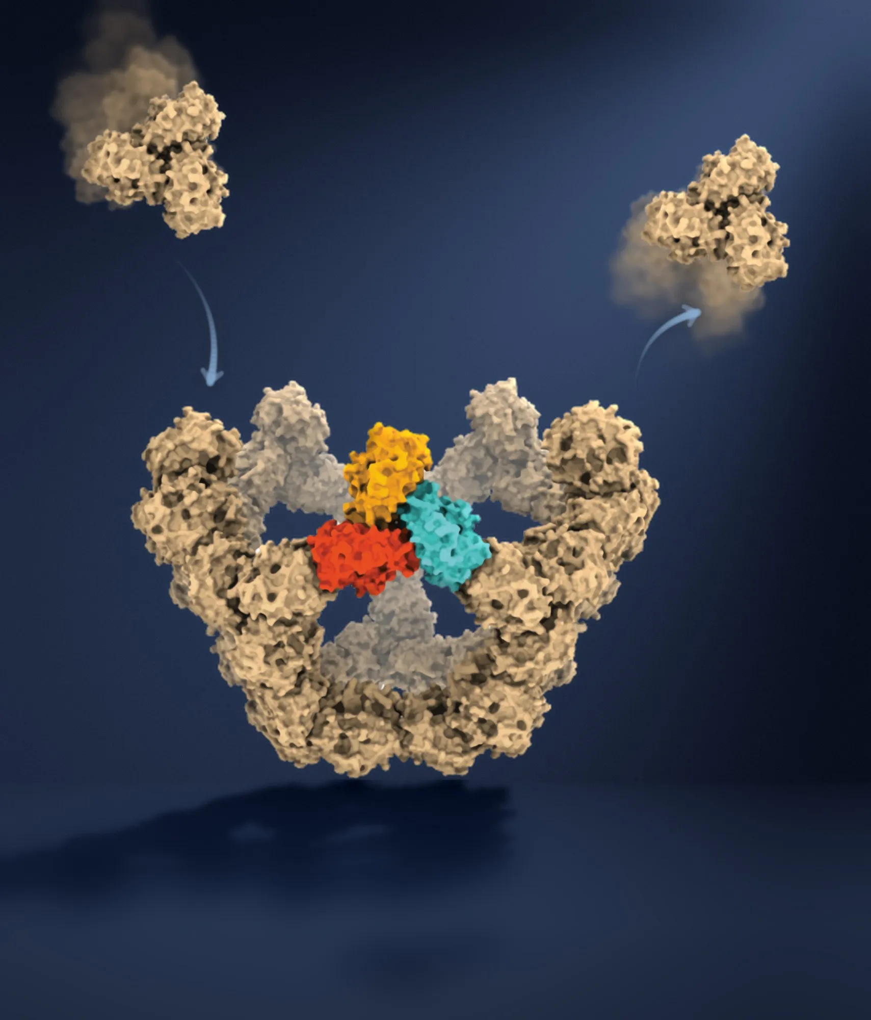
How the particle assembles: protein building blocks repeatedly join and leave; when they close a face, multiple connections lock them in place and assembly can continue from there. Structures rendered from PDB 7B3Y using ChimeraX; the illustration was subsequently edited using Claude (Anthropic), according to the University of Oxford. Illustration: Roi Asor / University of Oxford.
Vaccines, antivirals and other cell machinery
Co-author Jack Tan, of the MRC Weatherall Institute of Molecular Medicine, said understanding these processes at the molecular level “could have important applications in vaccine development and antivirals that disrupt viral assembly”. The researchers say the approach can also be used to study other self-assembling structures in cells, such as the cell’s internal skeleton, one molecule at a time.
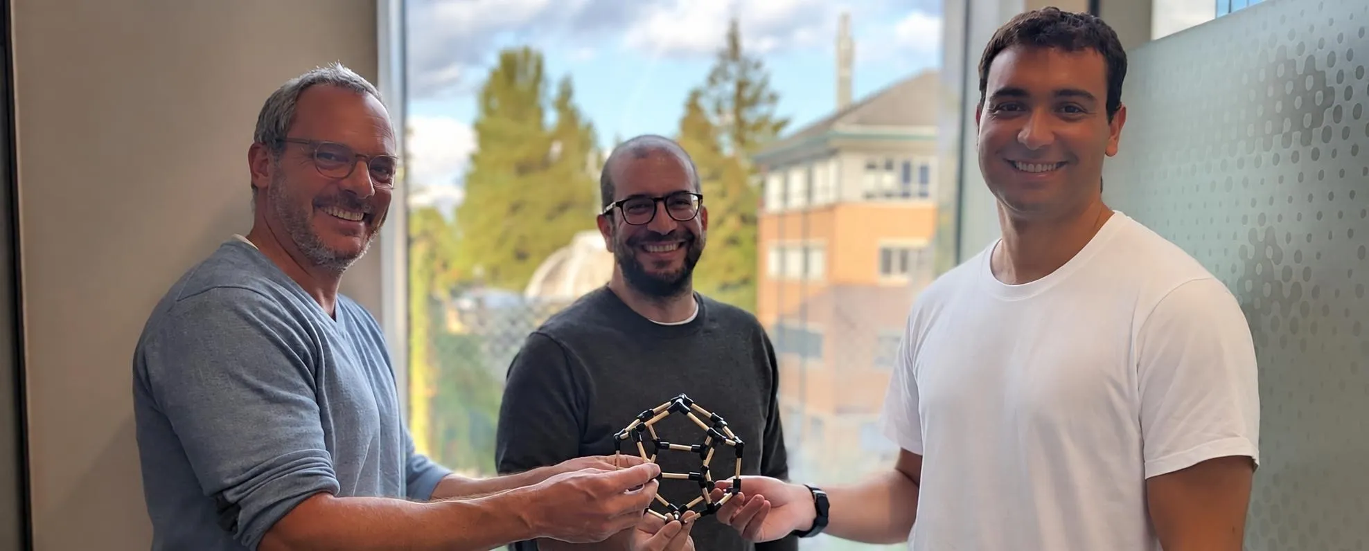
Photo and illustration courtesy of the University of Oxford.