Barcelona hospital to build digital twins of patients to test treatments before using them
Vall d'Hebron University Hospital will work with tech companies on virtual replicas of organs built from each patient's own data, to predict how their disease will progress and which treatment will suit them best. The project has €11.7 million in EU regional development funding and three years to run. It is only just starting: there are no results yet, and no companies have been chosen.
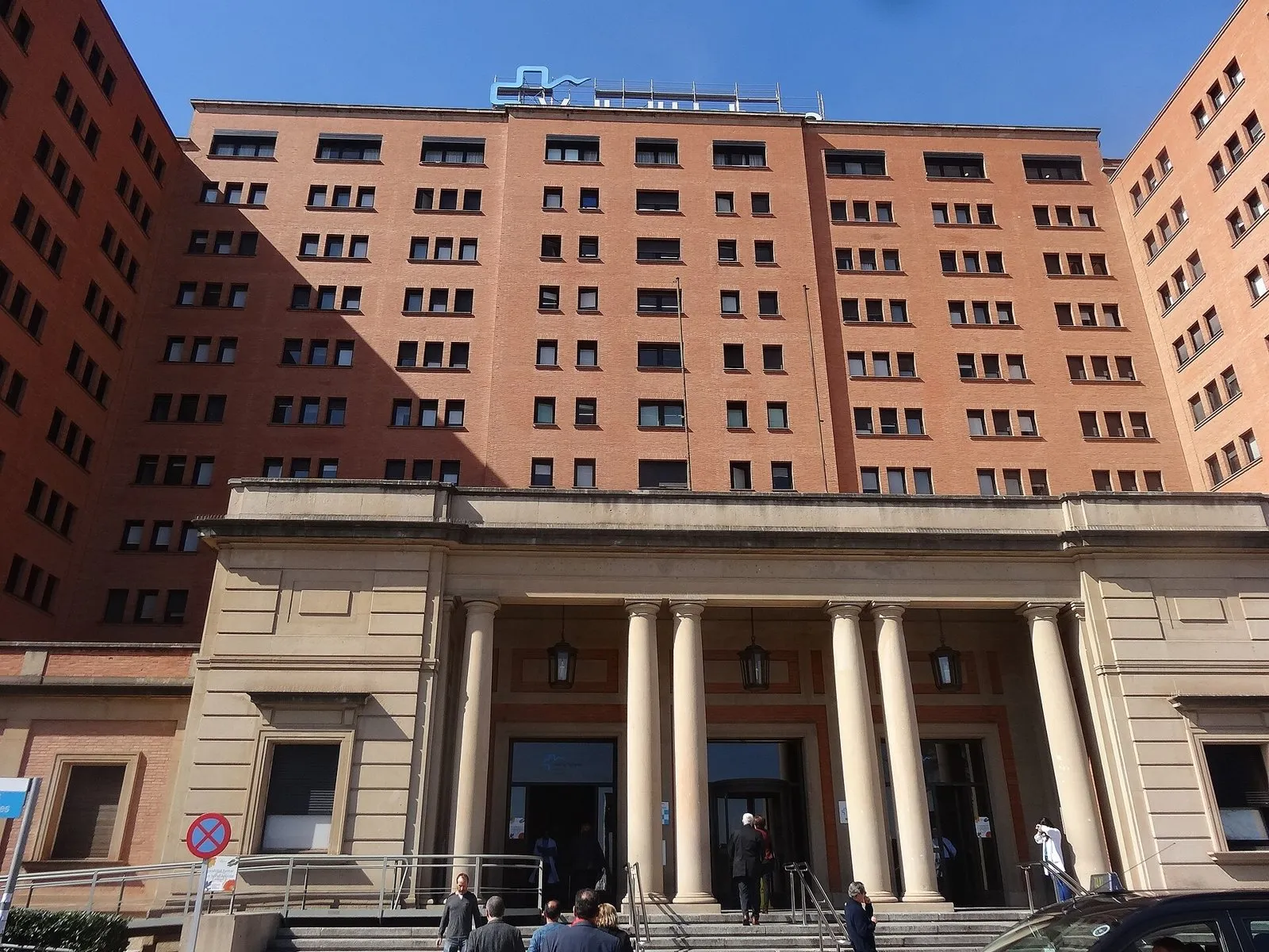
A virtual copy of a child’s heart on which doctors can rehearse a procedure before doing it for real. That is one of the ideas behind the human digital twins project that Vall d’Hebron University Hospital in Barcelona will develop over the next three years. According to the hospital, it will be the first in Europe to co-develop this technology with industry.
A digital twin is a virtual replica of an organ, a body system or a biological process, built from a real patient’s data. It combines medical imaging, clinical records, genomics, artificial intelligence and computer models to simulate how a disease will progress and how it will respond to a treatment before that treatment is given.
Rehearse on the replica, not the patient
“Digital twins have predictive power: they anticipate how a patient will evolve or what effects a treatment may have before it is applied in the real world,” says Dr Manel Escobar, clinical director of radiology at the hospital. They “must support clinical decisions,” he adds.
The starting point is the hospital’s 3D unit, which already builds personalised anatomical models to plan complex operations. The next step is for those models not only to show anatomy but to simulate how the organ works and how the disease advances.
From children’s hearts to migraine
The programme, called 3Dx3C, brings together ten strands of work. They include digital twins of the electrical activity of children’s hearts, early detection of heart disease from electrocardiograms and treatment for migraine. There will also be models to plan ablation for ventricular tachycardia, percutaneous pulmonary valve implants, the removal of liver tumours and surgery for tetralogy of Fallot, a congenital heart defect. The 3D models will be viewable in the operating theatre, including in augmented reality.
The project has €11.7 million from Catalonia’s share of the EU’s European Regional Development Fund, under the Strategic Technologies for Europe Platform (STEP). According to the hospital, it is the only health project among the eight STEP projects in the Catalan programme.
What is still to come
Vall d’Hebron still has to sign agreements with the tech companies that will build the models, and there are no results yet. According to Redacción Médica, once this three-year phase ends, models trained on the hospital’s patients will be validated in studies with patients at other centres.