Spanish study in Nature finds a back-up route for blood vessel growth
Rui Benedito's team at Spain's CNIC and Germany's Max Planck Institute shows that blood vessels can keep growing without VEGF-VEGFR2 signalling, long thought essential. It may help explain why some tumours stop responding to anti-angiogenic drugs. This is basic research in animal models: there is no treatment or patient trial yet.
For years, VEGF-VEGFR2 signalling has been seen as the master switch of angiogenesis, the process by which the body builds new blood vessels. That is why many cancer drugs block it: no vessels, no blood supply for the tumour. A study in Nature involving Spain’s National Centre for Cardiovascular Research (CNIC) shows vessels can grow even without it.
The caveat matters: this is basic research in animal models, followed through embryonic development and after birth. It points to new targets, but there is no treatment yet.
Cells that turn “venous”
Rui Benedito, who heads the Molecular Genetics of Angiogenesis group at CNIC and a department at the Max Planck Institute for Molecular Biomedicine in Germany, and his team built a genetic mosaic system to track normal cells and cells lacking VEGFR2 side by side. “This allowed us to observe how both cell populations behave throughout embryonic development and after birth,” says Irene García González, a CNIC researcher and co-first author.
Losing the receptor mainly hit the first phase of growth, and not equally in every organ: brain vessels held up well, while those in the heart, kidney and liver were far more sensitive, according to CNIC.
Over time, the cells without VEGFR2 rejoined the network, especially in veins and the small vessels around them. They did so by taking on the genetic features of vein cells, a shift the authors call venousization. Two other receptors, VEGFR1 and VEGFR3, helped them survive and migrate.
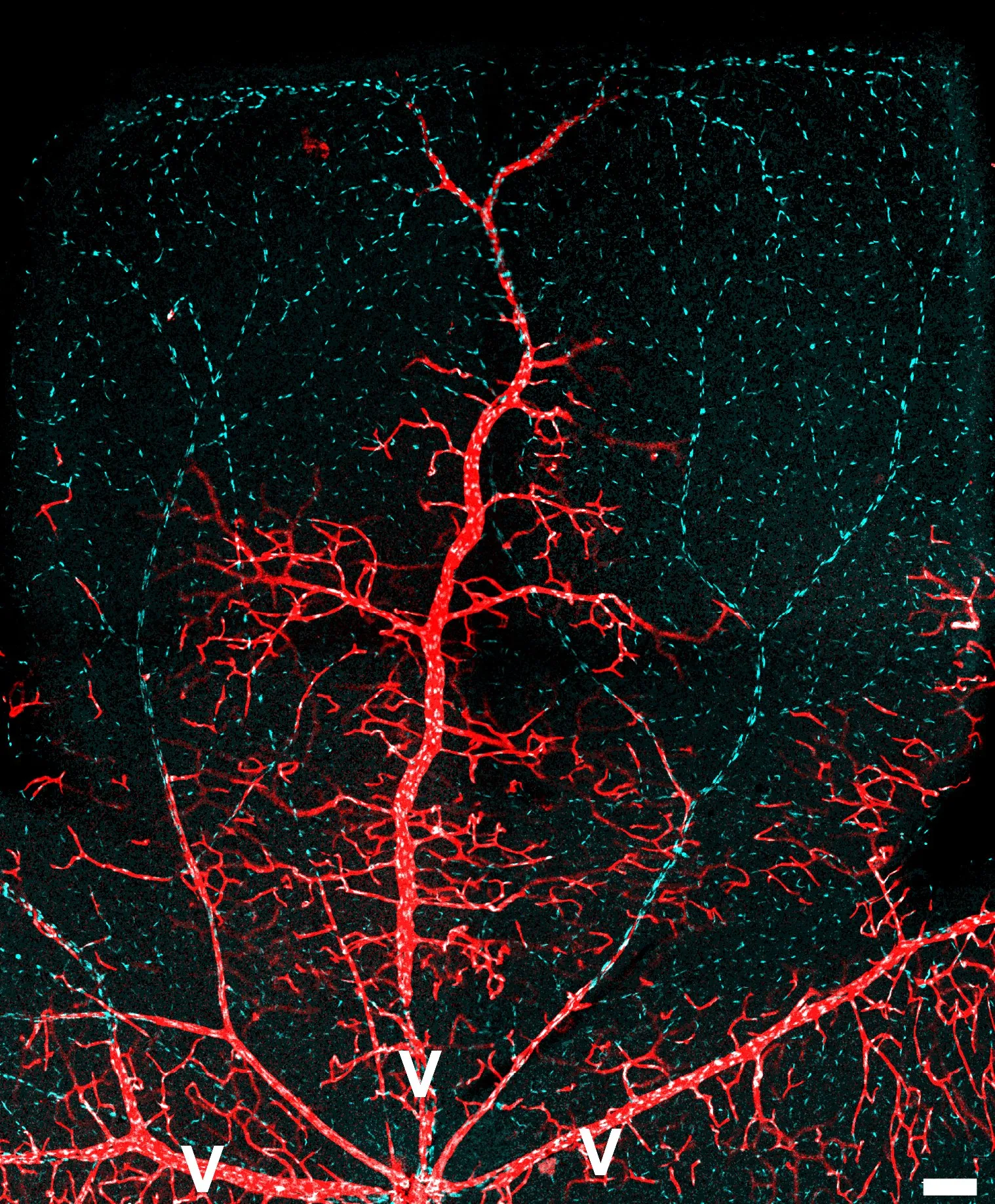
Blood vessels in a retina, in a microscopy image from the study. Image courtesy of CNIC.
A clue to treatment resistance
“These results may also help explain why some tumours become resistant to treatments that block VEGF-VEGFR2 signalling,” says Benedito, the study’s co-lead.
According to CNIC, the finding suggests that targeting that pathway alone may not be enough for lasting responses, and points to studying combination therapies that also block the back-up routes.
The microscopy was done at CNIC’s Microscopy and Dynamic Imaging Unit. Funding came from the European Research Council, Spain’s science ministry, the “la Caixa” Foundation and bodies in Sweden and Finland.